Hospital Enrique Tornú, City of Buenos Aires, Argentina
Abstract
Objective
To establish the frequency of complications in the different pathologies, differentiate them according to the type of stent used, and relate them to the time the different prostheses remain in place. To determine the average times in place of the prostheses in the cure of benign stenosis.
Material and method
Retrospective analysis of 388 interventional bronchoscopy reports performed to treat 300 patients with total or partial airway obstruction, due to lesions of benign or malignant nature. Therapeutic success was evaluated according to its capacity to restore the tracheobronchial lumen.
Results
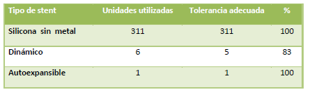
300 patients with partial or total airway obstruction, 115 women, mean age 52 ± 16.26, range 14-86 years. 388 therapeutic procedures were performed and airway recanalization was achieved in 96.33% of cases. A total of 318 prostheses were used, with a high general tolerance to stents of 99.68% and of 100% for the metal-free silicone prostheses. In 126 patients with classic post-intubation benign stenosis, 107 prostheses were used for 122 tracheal and 4 bronchial stenoses. The cure rate was 44%. In those in which prostheses were used, these remained installed for a period of 21.2 ± 13.30 months for the cured cases. The most frequent complication in the treatment of benign stenoses was stent migration: 13%, greater in straight silicone prostheses (85.7%) compared with the stenotic ones (14.3%). The development of granulomas (9.24%) was more frequent with the use of straight models (60%).
The second group brings together 174 patients with "non-benign-stenosis" conditions, containing 110 cases of carcinoma, 10 identified endobronchial metastases of extrapulmonary tumors, and the rest corresponding to various etiologies. 211 prostheses were used in this group, resulting in an average of 1.2 ± 0.46 stents per patient. The complication rate was 6.32%, with hemorrhage in first place. The mortality attributable to the treatment, in the 388 procedures performed, was 0.25%.
Conclusions
Rigid bronchoscopy shows its capacity to restore the lumen in situations of airway obstruction, with a success rate above 96% and very low mortality. Silicone prostheses with "stenotic"-type designs are decidedly more effective than straight models for the treatment of benign tracheal stenoses. Although the time in place necessary for the cure of benign stenoses has not yet been defined, this period may lie around 22 months when a stenotic model is used, but greater when a straight stent is applied.
Introduction
The endoscopic treatment of lesions that produce occlusion or subocclusion of different magnitude in the trachea, main bronchi or some lobar bronchi has achieved sufficient diffusion within the international medical community, to the point that its performance is well established (1). The indication to apply a method that can recover the pulmonary ventilation suspended by the presence of an obstructive lesion arises immediately in the bronchoscopist. However, it has remained limited to those cases in which open exeresis treatment is not possible. The use of silicone endoprostheses is another widely used resource in order to provide additional support to the airway after treatment and to prolong its effectiveness. Thus, endosurgical treatment has been simplified and interventional physicians have accumulated experience in its performance, a task that has shown to provide benefits proportional to the magnitude of the difficulties overcome during its implementation (2-3).
Patients and method
Data were obtained from interventional bronchoscopy reports corresponding to 388 procedures performed to treat 300 patients, with benign and malignant conditions that partially or completely occluded the large airway.
The interventionism applied to the patients was performed in one or several sessions. None of them was amenable to resection treatment by conventional surgery, and all underwent a flexible bronchoscopy in order to evaluate the characteristics of the lesion. Thus, the indication for the procedure was maintained considering the possibility of pulmonary re-expansion and the clinical presumption of improving the dyspnea with treatment, in all cases of malignant disease and in benign stenoses that reduced the lumen by 50% or more. This subjective evaluation was established by comparing the diameter in the affected area with that existing in the healthy airway near the lesion, and with the reference of the known caliber of the bronchoscope in use.
The patients underwent treatment with a rigid or flexible bronchoscope, under general anesthesia and muscle relaxation, in the operating room and under continuous control of cardiac and respiratory function. They received 2 grams of intravenous cephalothin in rapid injection minutes before the procedure.
The treatments were carried out by two experienced pulmonologists, with exclusive dedication to bronchoscopy.
For airway recanalization, dilation maneuvers were performed with bougies of increasing diameter, balloon and/or rigid bronchoscope. An electrocautery was also used to make cuts in some benign stenoses and thermocoagulation with tissue vaporization in malignant lesions. The cases, for the most part, were treated using a rigid bronchoscope with interchangeable tubes of different calibers, a set of optics, prosthesis introducers, forceps suitable for their mobilization, probes and tapes for the application of Montgomery-type T-tubes. In a few cases, the treatment was performed or completed with an Olympus BF1T30 flexible bronchoscope.
The application of different models and dimensions of silicone airway stents was arranged, although dynamic and self-expandable stents were also used. "Good tolerance" to the stent was accepted as the absence of clinical manifestations that would motivate its removal.
The result was considered based on having achieved sufficient recovery of the airway lumen.
In the benign stenoses, it was established to determine as satisfactory tracheal or bronchial recanalization all those in which the lumen of the affected area had reached 75% or more of the normal caliber for that case, after the end of treatment.
All those occurring during or after treatment were considered complications when they were directly linked to it. Thus, hemorrhage, desaturation with a decrease greater than 4%, rupture of the airway wall, restenosis, stent migration, development of granulomas and prosthesis colonization are recorded.
Cases of death were recorded when it was due to the instituted treatment or a direct consequence of it, during its application or afterward.
It was established to consider "cured" all those patients who, treated for benign stenosis and after removal of the stent when one had been implanted, remained free of recurrence and with an airway lumen greater than that they had before treatment, and who at the same time did not present dyspnea or stridor when carrying out their usual daily tasks.
Results
388 procedures were performed in 300 patients (1.29 procedures per patient).
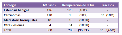
The procedures performed with a rigid bronchoscope totaled 383 (98.7%), while in 5 cases the flexible bronchoscope was used (1.3%). In 11 cases out of the 300, recovery of the bronchial lumen was not possible and it was then considered a failure of the procedure. All of them corresponded to patients with a diagnosis of carcinoma, and the endobronchial tumor lesion was also intramural and infiltrating. Airway recanalization was possible in 289 patients (96.33%). Table A.
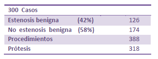
Of the total, 115 were women (38.33%) and 185 men (61.66%), in an age range of 14 to 86 years. The mean age was 52 years ± 16.26 and, for the treatment, 311 silicone prostheses (97.8%), one polyflex® self-expandable stent and 6 Freitag dynamic prostheses were used, adding up to a total of 318 devices. In this way, 126 benign stenoses in the trachea and bronchi (42%) and 174 "non-benign-stenosis" conditions (58%) were treated, Tables B-C. This last group comprises 110 cases of carcinomas that include 10 metastases of extrapulmonary primary tumors.
As for the use of prostheses by pathology, the 126 benign stenoses required 107 stents for their treatment (0.84 stents per patient).
For the group of "non-benign-stenosis" lesions, of 174 cases, 211 devices were used (1.2 ± 4.6 prostheses per patient). Table D.
In the 388 procedures, complications occurred in 44 of them (11.34%), which will be detailed below.


Benign stenosis group
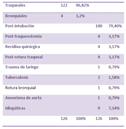
Of the 126 cases with benign stenosis, 122 affected the trachea (96.82%) and 4 (3.18%) the main bronchi. 100 of the total (79.40%) were post-intubation stenoses. Of the remaining 26, 4 occurred at the site of a previous tracheostomy, 4 were recurrences at the site of an end-to-end anastomosis from open surgery previously performed for the resection of the tracheal stenosis, 4 following tracheal rupture, one after severe laryngeal trauma and another due to aortic aneurysm. The cause could not be determined in 9 cases.
Four benign stenotic lesions were in the main bronchi: two were sequelae of pulmonary tuberculosis, one following traumatic bronchial injury and another following anesthetic bronchial intubation.
The tracheal stenoses were of complex conformation in 113 cases (89.7%), simple in 11 (8.7%), and 2 were subglottic (1.6%). Table E.
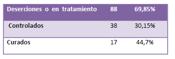
The controlled follow-up of the patients was extremely low. 88 cases (69.8%) did not attend the established appointments. Although many cases of this group are still in treatment, a large number have lost contact with the endoscopy service completely, including also those whose stent had been removed because the sufficient implantation time was considered to have ended.
Of the remaining 38, 17 cases achieved cure (44.7%) and 21 required a permanent stent, some additional treatment such as definitive tracheotomy, or continue in treatment. Table F.
The minimum time in place of the prosthesis was one month and the maximum known was 106 months, with a general average time in place of 19.6 months. Table G.

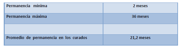
In the group of 17 cured patients, 11 prostheses were used. Each patient received one stent and 6 did not require any. The shortest time in place recorded is 2 months, for a "T"-tube; but for classic straight silicone stents, the lowest record indicates 10 months and the maximum 36, with an average time in place for the cure of 21.2 months. Table H.
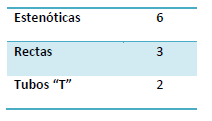
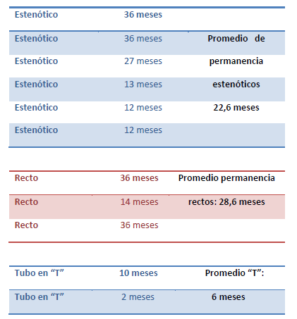
The 11 prostheses used by the cured patients were all silicone devices, a classic one-piece model without metal. Six of them stenotic, 3 straight and two "T"-tubes. Tables I-J. Finally, the average in months that the stents remained installed according to the models was 22.6 months for the stenotic ones, somewhat more (28.6 months) for the straight ones, and 6 months for the "T"-tubes. Table J.
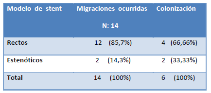
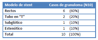
Complications: complications occurred in 44 of the 388 procedures (11.34%), all performed with a rigid bronchoscope. The five treatments instituted with a flexible bronchoscope had no complications. The greatest number of them occurred in the group of patients with benign stenosis, with a total of 33 in the 126 cases. The complications observed in the treatment of benign stenoses were mostly related to the 107 prostheses used. Fourteen (13%) were migrations, 6 colonizations (5.6%), development of granulomas in 10 cases (9.24%), mucous hypersecretion in 2 (1.86%), and one rupture of the posterior wall of the trachea (0.79%). Table K.

The migration and colonization of the prostheses was more common in straight silicone models and less frequent in the "stenotic"-type designs. Table L.
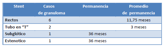
As for the development of granulomas by contact with the stent, occurring in 10 of the 107 prostheses used, they happened on six occasions in which a straight stent had been applied, two with a "T"-tube, one with a subglottic stent and another with a stenotic one. The general time in place for all these prostheses was 17 months; but individually, the complication was detected on a subglottic stent applied for 36 months, a stenotic one for an identical period, and, as a group, 3 months for the "T"-tubes and, finally, the straight stents that generated granulomas had an average time in place of 11.75 months. Tables M-N.
"Non-benign-stenosis" group
The group of patients not included with the benign stenoses is made up of all the cases that presented other invasive lesions of the tracheobronchial lumen or that developed their growth in it; and although it is composed in greater number of bronchial carcinomas, other etiologies were also found in this set, and none of them, as mentioned, can be included in any way with the benign narrowings that affect the airway.
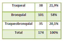
Thus, this group includes 174 patients, of whom 38 had their condition located in the trachea, 101 in the bronchi and 35 shared both locations. Table O.
In 11 (10%) of the 110 cases of carcinoma, reconstruction of the tracheal or bronchial lumen was not possible. Table A.
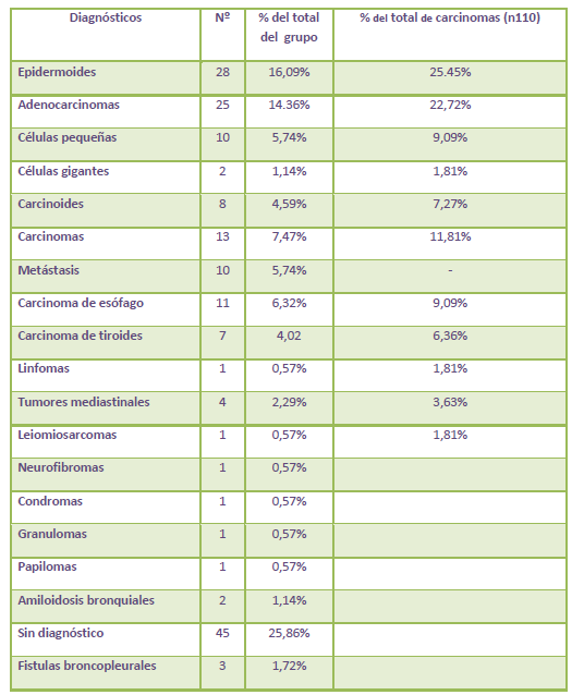
As for the histological type of the lesions, 28 epidermoid carcinomas, 25 adenocarcinomas, 10 small-cell carcinomas, 2 giant-cell carcinomas and 8 carcinoid tumors were found. The endobronchial metastases totaled 10 cases. In another 13, the endoscopic biopsy identified the carcinoma but was not sufficient to establish its histology.
In addition, 1 chondroma, 1 neurofibroma, 11 esophageal carcinomas that invaded the bronchial, tracheal or both lumens; 7 thyroid carcinomas involving the tracheal wall, 4 mediastinal tumors and 1 lymphoma were found. In one case the diagnosis was papilloma, in another leiomyosarcoma, 1 granuloma and 2 bronchial amyloidoses. In a further 45 cases the diagnosis could not be established by flexible bronchoscopy, and they were equally subjected to endoscopic disobstruction treatment. Epidermoid carcinoma was the most frequent tumor with 25.45% of cases, followed by adenocarcinoma (22.72%) and oat cell (9.09%). Three patients with bronchopleural fistula also received bronchoscopic treatment, in whom the fistula was blocked with a cylindrical, solid silicone device. (Table P.) In this group, 211 prostheses were used for 174 patients. (Table D).

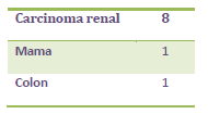
In relation to the 10 metastases, 8 of them were from clear-cell renal carcinoma, one from a breast tumor and the other from the colon. Table Q.
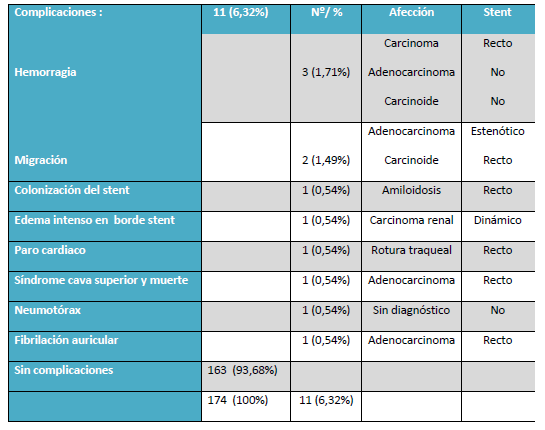
Complications occurred in 11 cases (6.32%), led by hemorrhages that equaled the suction capacity of the aspiration system, 3 in total (1.71%), which resulted from the treatment of a non-typified carcinoma, an adenocarcinoma and a carcinoid tumor.
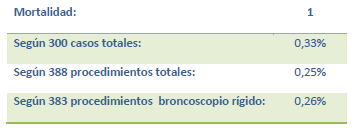
Migration of the prosthesis in two cases (1.49%): tracheal adenocarcinoma with a stenotic stent in the first, and a straight stent in a carcinoid tumor, the second. Colonization (0.54%) of a straight prosthesis implanted in a case of amyloidosis. The list of complications of the present "non-benign-stenosis" group of lesions continues with a case of intense mucosal edema at the end of a dynamic stent (0.54%), reversible cardiac arrest during the treatment of a tracheal rupture (0.54%), a superior vena cava syndrome (0.54%) followed by death at 48 hours in a patient with adenocarcinoma that invaded the trachea and both main bronchi (0.54%), an atrial fibrillation in a case with adenocarcinoma (0.54%) and a partial pneumothorax in a patient lacking histopathological diagnosis. Table R. Thus, the mortality determined for this group of 300 patients is 0.33%, and considering the mortality linked to the procedure, it is reduced somewhat more, to 0.25%. Table S.
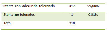
Intolerance to the prosthesis turned out to be extremely rare. It was only observed in one case in the entire series, representing 0.33% in relation to the treated patients and somewhat less, 0.31%, if considered in terms of the stents used. Tables T-U.
It is a dynamic "Y" prosthesis implanted in a patient with intrabronchial metastasis of a primary clear-cell renal tumor. The intolerance, manifested by intractable coughing, was attributed to the intimate pressure contact of the proximal end of the prosthesis with the tracheal mucosa adjacent to one of its rings. The stent was shortened and reinstalled without symptomatic relief and had to be removed and replaced.
Discussion
The usefulness of the method is well consolidated and the selection of patients has been widely commented on and published. Thus presented, the debate on these points would be, given its lack of novelty and the even smaller contribution of knowledge, monotonous and unnecessary. Observing the frequencies of appearance of problems, or the lack of them, considerations arise that are the start of this discussion. The series of 300 cases contains a numerous and exclusive group of benign stenosis, mostly tracheal. In all of them the caliber of the airway was recovered with endoscopic interventionism. So the procedure in general was clearly satisfactory, with a success in achieving its purpose of 96.33%.
Having announced electrocautery as the only cutting, vaporization and thermocoagulation device used in this series, its resolution capabilities bear an obligatory relation to the 96.33% of resolved cases (4-6). The 3.66% of failures corresponds to 11 cases with diagnoses of carcinoma. In them, the procedure fails to find the lumen distal to the airway obstruction (5), as happens when the lesions turn out to be intramural and highly infiltrating, that is, when this sought-after lumen does not exist.
The overwhelmingly superior use of classic silicone prostheses is due to their simple availability in our setting and, safeguarding their well-known behavioral virtues, their preference is not based on the detriment of other types and designs, which are also efficient (7-9-10).
The dropout of operated patients is strikingly high. Adding the cases that are in follow-up or in treatment at the time of preparing this account, they reach 69.85% of the total of benign stenoses. Thus, better general conclusions could not be obtained about all the points examined here. Equally, the time the stents have remained implanted in the patients has been observed. There are few reports about the time prostheses remain in place. In the reviewed literature, the devices were removed due to cure, remission of the treated lesions, or simply removed after a period considered sufficient for stabilization or cure (11-12), without the results of the long therapeutic process being known in all of them. In many cases the device remained implanted throughout the patient's survival (8). They are usually kept in the airway for variable periods, from a few weeks to 24 months or more, depending on the patient's survival in malignant diseases or on the control of their condition. This varies according to the therapeutic preferences of the work groups. There are reports of very prolonged stays, which on some occasions have exceeded 4 and 5 years, with good tolerance (13). Removal of the device for other reasons such as its deterioration, modification, decrease or loss of its mechanical qualities, or rejection of its components, was not observed.
In the series presented, the devices remained installed for a range as wide as 1 to 106 months, depending naturally on very different circumstances, such as the individual behavior of each case, its operability, evolution and, precisely, the prosthetic element selected. The "T"-tubes are used for a short period, since they usually fulfill an intermediate "step" on the path of the patient's therapeutics; whereas the stents remain for a much longer period, since they attempt to offer the tracheal or bronchial wall support during the time required for its consolidation, since what only comes with time cannot be obtained in less.
We have examined the time in place, relating it to the etiology, and likewise the time in place of the prostheses in the cured benign stenoses, which was, both on average and considering each prosthesis individually, shorter for the stenotic-model silicone stents (22.6 months) than for the straight ones (28.6 months). For this fact of full certainty to acquire the force of truth, it will be necessary to study a more numerous group of cases, but it could be indicating a trend.
The stenotic prostheses also showed less migration. Although this situation is to some extent expectable, since it has a profile that tries to imitate the contour of the stenosis in order to hinder its displacement, which is, in short, the reason for its design; and with its choice we see our purpose fulfilled. The stenotic stents showed less colonization, though this time we cannot offer a thorough basis to explain it. The granulomas were present, as they always are. This time with a frequency of 9.4%. The granulomas should not occur if the stents were correctly applied, that is, with the middle portion of the prosthesis "fitted" in the stenosis and its ends "floating" in the tracheal lumen.
The analysis of the "non-benign-stenosis" group revealed, first, a greater quantity of prostheses used (211 units), a cause found in the need to use more than one stent per patient and also sometimes more than one procedure. This in turn indirectly shows that the probable extension of survival allows the malignant disease to progress and, with it, to recur by obstructing the airway at the same or a different site, motivating a new intervention.
They mostly correspond to bronchopulmonary carcinomas or those of neighboring organs. Among the latter, that of the thyroid, with locoregional extension and invasion of the tracheal lumen, a circumstance and etiology that in no way modifies the bronchoscopist's attitude. Eleven cases of esophageal carcinoma which, unlike the previous one, damages and breaks through the posterior wall of the trachea or, when its location is lower, involves the left main bronchus, again on its posterior wall, since it is there that the esophageal path crosses this bronchus. Often the damage is of left para-carinal location, with the esophagus coming to inhabit the bronchial lumen. It has been very useful, to endoscopically realign the left main bronchus, expel the esophagus and cover the fistulas that are a frequent companion in this picture, to use a smooth-walled silicone stent, since anchors are not necessary now. One of its ends widens in the shape of a cone and coincides anatomically with the origin of the main bronchus. The beveled distal end makes the maneuver of entering the bronchus and introducing the stent simple.
Migration of the prosthesis, although less common (11) in malignant conditions, is also possible. Only two migrations (1.49%) are described here, one of which corresponds to a carcinoid tumor in which an error in the early typing of the lesion led to the treatment of the obstruction with stent implantation.
Few comments will be made about two fortunately infrequent facts, such as intolerance to the stent, whose reasons have already been clearly developed; and death as a result of the procedure, which in our report was 0.25%. It corresponded to a patient with mediastinal disease due to an adenocarcinoma. Since the mediastinal syndrome makes rigid bronchoscopy risky for diagnostic purposes, this risk will be reasonably increased when the procedure is also therapeutic, since the same factors that operate in the first will be more influential in the second, because the time necessary for the intervention will be, for easily understandable reasons, much greater. Thus, the maneuvers it entails and the displacement of tissues caused by the rigid bronchoscope lead to an increase in local inflammation and a worsening of the already compromised venous return to the mediastinum. Such was the case in our cited case.
This work was intended to be a recount of the results regarding what happened after the interventional assistance of a moderately numerous group; however, it took a turn in its meaning upon observing figures that seem to mark a trend regarding the possible relationship between the use of prostheses and their complications linked to the time in place, revealing that, at least for this series, the time in place of a stent required for the cure of benign stenoses can be situated "around 23 months". The case series is not sufficient to grant it the character of a firm recommendation, but a timid orienting usefulness is noticed. Thus, the lines of a direction to follow are traced, toward the encounter of a solution that possesses the magnitude of the effort hitherto committed to stopping this condition, so simple and rebellious at the same time.
References
- Cavaliere S, Venuta F, Foccoli P, Tonielli C, La Face B. Endoscopic treatment of malignant airway obstructions in 2008 patients. Chest 1996; 110: 1536-42.
- Stephens K E, Wood DE. Bronchoscopic management of central airway obstruction. J Torac Cardiovasc Surg. 2000;119:289-96.
- Dumon JF, Dumon MC. Dumon-Novatech Y-Stents: A Four-Year Experience with 50 tracheobronchial Tumors Involving the Carina. J Bronchol 2000; 7:26-32.
- Sutedja, T. Endobronchial electrocautery is an excellent alternative for Nd-YAG laser to treat airway Tumors. J of Bronchology 1997;4:101-105
- Boelcskei PL, Dierkesmann R, Bauer PC, Becker HD, Bolliger CT, Wolfgang FJ. Section on respiratory endoscopy of the German Society of Pulmonology. Recommendations for bronchoscopic treatment of tracheobronchial occlusions, stenoses, and mural malignant tumors. J Bronchol. 2000;7:133-8.
- Cynthia Huisman, Klaas W. van Kralingen, Pieter E. Postmus, Tom G. Sutedja. Endobronchial Lipoma: A Series of Three Cases and the Role of Electrocautery. Department of Pulmonology, Academic Hospital Vrije Universiteit, Amsterdam, The Netherlands. Respiration 2000;67:689-692
- Teruomi Miyazawa, MD, FCCP, Michio Yamakido, MD, FCCP, Sadao Ikeda, MD FCCP, Kinya Furukawa, MD, Yuichi Takiguchi, MD, Hirohito Tada, MD, and Takayuki Shirakusa, MD. Implantation of Ultraflex Nitinol Stents in Malignant Tracheobronchial Stenoses. Chest October 2000 vol. 118 no. 4 959-965
- Jantz, MA, Silvestri, GA. Controversy Silicone Stents versus Metal Stents for Management of Benign Tracheobronchial Disease. J Bronchol 2000; 7:177-183.
- A. Cosano Povedano, L. Muñoz Cabrera, F.J. Cosano Povedano, J. Rubio Sánchez, N. Pascual y A. Martínez Escribano Dueñas. Cinco años de experiencia en el tratamiento endoscópico de las estenosis de la vía aérea principal. Arch Bronconeumol. 2005;41 (6):322-7
- Marc Noppen, MD, PhD, FCCP; Grigoris Stratakos, MD; Jan D'Haese, MD; Marc Meysman, MD, FCCP; and Walter Vinken, MD, PhD. Removal of Covered Self-Expandable Metallic Airway Stents in Benign Disorders: Indications, Technique, and Outcomes. Chest 2005; 127:482–487.
- A. Brichet, P. Ramon, C.H. Marquette. Sténoses et complications trachéales postintubation. Réanimation 2002; 11: 1-10