Small-caliber biocompatible silicone bronchial stent, specifically designed for the common trunk of the basal segments of the lower lobes. Its conical anatomical shape mirrors the taper of the bronchus, avoiding the distortion caused by conventional cylindrical stents.
Product code: ROLL


Code ROLL
The Stening® Roll 911 —also known as the ROLL stent or small-caliber bronchial stent— is Stening’s response to the clinical need to treat conditions of small bronchi that are still able to provide ventilation to their distal segments, as occurs with neoplastic infiltrations in the right and left lower lobe bronchi.
It is a special stent for the common trunk of the basal segments of the lower lobes. Its smaller, more widely distributed anchors provide gentle support; its anatomical shape mirrors the taper of the bronchus, avoiding the distortion caused by conventional cylindrical stents. The preferred size, the ROLL 911, has an external distal diameter of 9 mm and a proximal diameter of 11 mm, with a length of 20 mm. It is also supplied in two alternative sizes: the ROLL 810 (20 mm) and the ROLL 1012 (25 mm), the latter specifically for the bronchus intermedius.
Review the indications, available dimensions, instructions for use, care and warnings of the Stening® Roll 911.
The Stening® Roll 911 is indicated for the treatment of obstructions in small-caliber bronchi that retain ventilation of their distal segments.
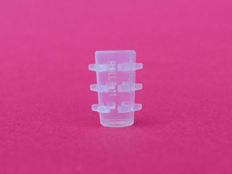
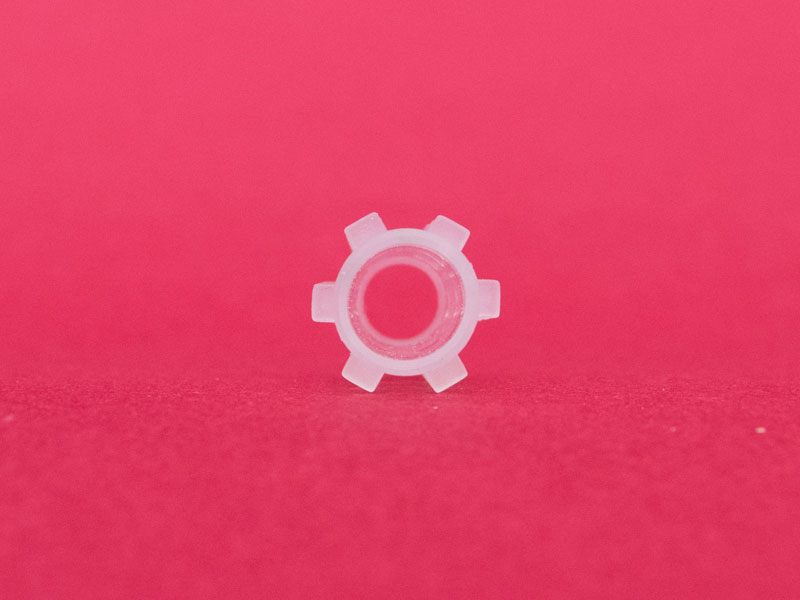
The Stening® Roll is offered in three sizes with differentiated external distal and proximal diameters, which reproduce the taper of the bronchus to be treated.


Measurements in millimeters. The distal (A) and proximal (B) diameters correspond to the ends of the stent and the length (C) to its total span. For specific inquiries about instruments, bronchoscopes or introducers, contact us at (+54) 11 4553-5070 or (+54) 11 4551-2333.
The procedure is performed under general anesthesia. The implant of this type of prosthesis can be carried out directly through the working channel of the tracheoscope or bronchoscope, or by using a conventional introducer for silicone prostheses. The airway is accessed with a rigid endoscope.
The length and diameter of the area to be covered with the stent must be properly established. A simple method to determine the length of the affected area is to mark the tracheoscope when its tip reaches the end of the lesion, and to repeat the marking after withdrawing it back to the beginning of the lesion. The diameter of the trachea or bronchus must be estimated by comparison with the known diameter of the endoscope used.
The prosthesis is thus released. If necessary, it can be repositioned with an alligator forceps, the maneuver being simpler if the stent is placed “below” the lesion.
Steps 1, 2 and 3 are repeated. Then the tracheoscope containing the introducer and the prosthesis is stopped 5 mm before the lesion to be treated, and the ejector plunger is pressed slowly. In this way, the prosthesis will be expelled toward the affected trachea.
Some stent loader models are not introduced inside the tracheoscope, but simply attached to it at its proximal end, from where the stent is propelled. For this purpose, the endoscope will have been stopped proximally or distally to the lesion as explained above, in order to push the prosthesis with the plunger provided with the endoscopic instrument. The stent will then travel through the entire interior of the tracheoscope until reaching the trachea. At this point, a sudden reduction in resistance to the pressure exerted on the plunger will be perceived, indicating that the stent has begun to exit the interior of the endoscope.
The stent may require additional maneuvers in order to correct or adjust its final position. It is preferable to correct a stent that has been installed beyond the desired position than the opposite, since it is highly inconvenient to advance a prosthesis that has been released “before” the affected area.
To move a stent in a proximal direction, it can be grasped by its edge and pulled gently. We strongly recommend, for its precision, a maneuver consisting of grasping the stent by its edge as mentioned, and then advancing with the direct-vision optics inside the stent until visualizing its final end. Then pull the forceps and you will see the stent ascend through the airway. Stop the traction when you consider the position to be optimal.
Intubation is performed with a tracheoscope or rigid bronchoscope as appropriate. Easy to remove, the silicone stent must be grasped by its edge with an alligator forceps with sufficient firmness. The forceps is rotated about 360° so that the stent folds into an omega shape and thus loses its radial resistance to compression. Then the forceps is pulled, extracting the prosthesis together with the tracheoscope.
The proximal end of the stent may be introduced inside the tracheoscope. With this maneuver, the vocal cords are protected during extraction. Other methods of implantation and removal are also possible depending on the operator’s experience and preferences.
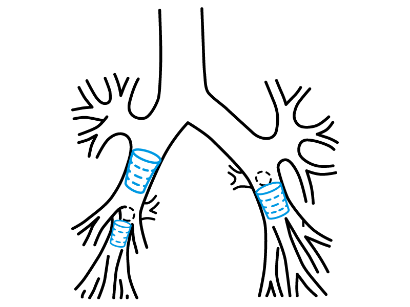
The division spurs of the basal segments stop the stent and prevent its implantation in an undesired site. For implantation, all sizes of the Stening® Roll can be introduced through a number 8 bronchoscope or one of larger diameter. Once the endoscope is positioned near the bronchus intermedius or lower lobe bronchus as the case may be, introduce the previously lubricated and folded Stening® Roll through the end of the bronchoscope, then push it with the bronchoscopy forceps until it leaves the endoscope at its opposite end and is thus lodged in the bronchus. A standard stent loader and its pusher for a number 8 or 9 bronchoscope, or whichever you prefer, may also be used.
When loading the stent into the bronchoscope, remember that the widest end of the Roll (letter “B” in the diagram) must be placed in the proximal position so that it faces the bronchoscopist. Conversely, the narrowest end will occupy the distal position. Removal is simple compared to that of a conventional stent owing to the small dimensions of the Stening® Roll.
The Stening® Roll can be trimmed at its end to fit the length of the bronchus in which it is implanted. Make the cut at its proximal end, since this end does not face any bronchial spur during respiratory dynamics. Implanting a stent in the common trunk of the lower lobe may result in occlusion of the entrance of the apical segment; the physician must weigh the benefits of restoring ventilation of the basal segments despite the loss of the apical segment of the lower lobe, when it is not already affected by the neoplastic disease.
Recommendations for follow-up of patients with a bronchial stent.
The device must not be reused. This prevents cross-contamination.
Contact us for personalized technical advice, size selection, custom manufacturing or inquiries about international shipping.